
How Does IPA-3 Suppress
the Cancer Growth in vivo Effectively?
Recently a group at University of Hong Kong (1) reported
that IPA-3, the direct inhibitor of PAK1-3 (group 1 of PAK family kinases) strongly
suppresses the growth of human hepatoma xenograft in mice even at very low dose
(4 mg/kg, i.p., three times a week). As far as my knowledge concerns, this is
the first report that IPA-3 works on cancers so effectively in vivo. However,
considering the IC50 in vitro (cell culture), which is around 30 micro M, it is
rather unlikely that the major (most sensitive) target of IPA-3 responsible for
the suppression of cancer growth in vivo is PAK1-3.
In a common sense, if a given drug ‘s IC50 in vitro
is around 30 micro M, the doses far more than 100 mg/kg would be required for
the suppression of cancer growth in vivo. In other words, something other than
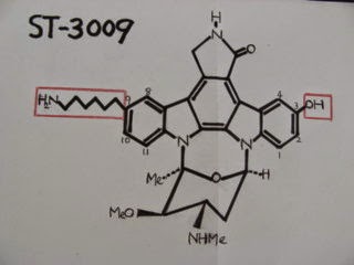
PAK1-3 could be the major target of IPA-3 or its metabolite in vivo. IPA-3
contains a disulphide bond which combines two 2-naphthalenol molecules, and in
cells it binds covalently the auto-inhibitory domain (AID) of PAK1-3, and
inhibits the binding to CDC42 at around 2 micro M, clearly indicating that its
cell permeability is pretty poor.
Generally speaking, the drug working at such a low
dose in vivo, its IC50 for the major target should be around 10 nM in cells, less than 1/1000 of the IPA-3’s
IC50 for inhibiting PAK1 in cells. Thus, it is most likely that IPA-3 is just a
“pro-drug”, and in vivo (circulation) it is converted to an unknown molecule
which then enters the cells far more efficiently, and inhibits its new (to be
identified) target at around 10 nM.
How can we identify this responsible metabolite of
IPA-3 and its target? Suppose we have a
radioactive S35-IPA-3 molecule in our hands. Inject this radioactive
molecule into mice or feed nematodes (C. elegans) with bacteria cultured in a
medium containing this radioactive molecule. Collect the serum from mice or an extract
of the whole nematode to analyze through HPLC. Before the analysis, the radioactive
serum or extract should be incubated with cancer cells, and select the
radioactive materials highly permeable through cell membranes, and determine
its major intracellular localization. If its binding to its target protein(s)
is covalent, we can identify the radioactive protein band (s) by SDS-PAGE.
Nevertheless, I suspect that the major target of
this IPA-3 metabolite would be PAK1-3 per se or among their major targets (effectors) such as LIM kinase and beta-catenin, because the phenotypes of both PAK1-deficient
(knock-out) and IPA-3-treated mice are very similar if not the exactly same.
Reference:
- Wong LL, Lam IP, Wong TY, Lai WL, Liu HF, Yeung LL, Ching YP. 2013. IPA-3 Inhibits the Growth of Liver Cancer Cells By Suppressing PAK1 and NF-κB Activation. PLoS One. 8: e68843.


![リオ五輪男子体操団体:日本(金)、ロシア[銀]、中国[銅]。](https://blogger.googleusercontent.com/img/b/R29vZ2xl/AVvXsEjHS61FORcH43CteZVfJzLmbqvNwOIliOSMpTpRtEi7x8j1ZwPk5rDaZovTrwuZxfDDtdEDSj673it735LF0mweIunaj7ja07lURBDYTV6wPMaAlumFt3aWWzYbHZgIaxcOLk_OKEMyQ3lX/s1600/2016+taiso+gold.jpg)
![皇太子(明仁)による沖縄訪問 [1975年]](https://blogger.googleusercontent.com/img/b/R29vZ2xl/AVvXsEjvSQrzV7yw_4gVQSwxZP_jh4VnEJscSqOqbiBh0VdAK3CRddXRqkd70JdLyws9fGejk-FGVmXWbHvSxlF3f8UogTyf9KXbqU1NGXesvcx2Hlsd6uq81AHweeioc61wynq3d2IYuyolijgT/s1600/akihito+message.jpg)


![アルニカ [ウサギ菊]](https://blogger.googleusercontent.com/img/b/R29vZ2xl/AVvXsEilqv0qou-4NpoUh1PFWYK0FSaozKazee0VYGxsFtfjBma46ya9yxqB6X9Ziuob25tNRpBbnFIcUFlOEjz1WcAjVNzjGl1E-QbDgE7VOLkjZDx0eplJ1WJHf0fTEWXxf8F5G-cHUhqHELY9/s1600/ArnicaS.jpg)


.jpg)


.jpg)


0 件のコメント:
コメントを投稿